LOCAL DRAFT · STAGING — NOT PUBLISHED

TG2 — the anti-TIGIT clone with documented advantages over TG1 in human FFPE IHC.

Clone TG2 · Cat. no. DIA-TG2-M · Distinct from clone TG1

Scientific USP

Documented advantages over TG1

Higher affinityTo TIGIT · vs TG1
Stronger tonsil stainingIHC intensity · vs TG1
Higher dilutabilityvs TG1

Relative statements vs TG1, tissue-bound (tonsil), not generalized (section P·06).

Mouse monoclonal antibody for studying human TIGIT in formalin-fixed, paraffin-embedded tissue. The clone-specific datasheet documents immunogen, detection-system-dependent starting dilutions, epitope retrieval, tonsil as positive control and a membranous lymphocyte staining pattern. TG2 shows higher affinity to TIGIT, stronger staining intensity in tonsil IHC and higher dilutability compared with TG1.

Evidence layer 01

Clone-specific datasheet

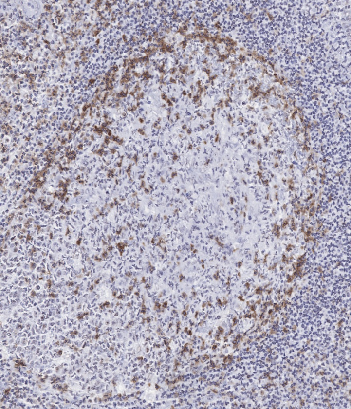
Datasheet DIA-TG2-M (V05, 25 Feb 2025; content identical to V03, 22 Feb 2022) documents identity, immunogen, formulation, reconstitution, storage, FFPE-IHC starting conditions, positive control and a reference image of human tonsil.

Evidence layer 02

Documented starting conditions

Heat-induced epitope retrieval in an autoclave (121 °C, 5 min, Tris-EDTA-citrate pH 7.8); 1:50 for biotin/(strept)avidin-based and 1:100–1:200 for polymer-based detection. A documented artefact note covers weak nuclear/nucleolar signal in some epithelia.

Evidence layer 03

Comparison with TG1

TG2 shows higher affinity to TIGIT, stronger staining intensity in tonsil IHC and higher dilutability compared with TG1 (section P·06).

TG2 reference image and historical TIGIT gallery: section P·03 ↓

P·02 Technical data

Controlled product data.

Product image of ONCOdianova anti-TIGIT antibody clone TG2, catalog number DIA-TG2-M
TG2 product image · Clone TG2 · DIA-TG2-M
ProductAnti-TIGIT (Human) from Mouse
CloneTG2 (monoclonal) — a separate hybridoma clone; not TG1
Catalog numberDIA-TG2-M
Distributor catalog numberODN-DIA-TG2-M (BIOZOL)
TargetTIGIT — T-cell immunoreceptor with Ig and ITIM domains (Gene ID 201633, UniProt Q495A1)
ImmunogenRecombinant peptide from the extracellular domain of human TIGIT (amino-acid region and epitope not published)
IsotypeMouse IgG1/κ (datasheet; the previous product page stated “IgG1” without light chain)
Host speciesMouse
ReactivityHuman
ConjugationUnconjugated
Format100 µl, lyophilized; antibody purified from culture supernatant
ReconstitutionRestore to 100 µl with sterile distilled water; gentle shaking for 10 minutes
FormulationPBS, pH 7.4, 2% BSA, 0.05% sodium azide
ApplicationsIHC on standard FFPE sections (datasheet V05)
IHC starting range1:50 – 1:200 — 1:50 for biotin/(strept)avidin-based detection, 1:100–1:200 for polymer-based detection; optimal dilution to be determined by the user for tissue, fixation, platform and detection system
Epitope retrievalHeat-induced epitope retrieval required; autoclave 121 °C, 5 min, Tris-EDTA-citrate pH 7.8 recommended
Positive controlTonsil
Staining patternMembranous (lymphocyte subsets)
Comparison with TG1Higher affinity to TIGIT, stronger staining intensity in tonsil IHC and higher dilutability compared with TG1 (relative statements, not generalized — section P·06)
StorageLyophilized at 2–8 °C; long-term at −20 °C (stable for at least one year); reconstituted at 2–8 °C short term (several weeks); avoid repeated freeze/thaw cycles
Associated antibodiesDIA-TG1-M (anti-TIGIT, clone TG1) · DIA-R12 (anti-PVRIG/CD112R, clone R12) — as listed in the datasheet; each clone carries its own evidence
StatusResearch Use Only

The technical specifications above are documented in datasheet DIA-TG2-M V05 (25 Feb 2025), whose product content is identical to V03 (22 Feb 2022); gene and protein entries link to their registries. No specification, protocol or publication of clone TG1 has been transferred to this table. Each laboratory must validate its own conditions.

P·04 Documented starting protocol

Datasheet procedure.

Only the TG2 datasheet applies. The procedure below is the “Instructions for Use” section of datasheet DIA-TG2-M V05 (25 Feb 2025; identical wording in V03). The previous TG2 page linked the tech note “TIGIT staining manual + automated” and showed a manual and a Leica Bond RX protocol – both are explicitly documented for clone TG1 there and are not TG2 protocols. No TG2-specific automated-stainer protocol is documented. Dilutions are starting values; each laboratory validates tissue, fixation, platform and detection itself.

ProcedureImmunohistochemical staining of standard formalin-fixed paraffin sectionsDatasheet DIA-TG2-M · V05 · 25 Feb 2025

Preparation and epitope retrieval

  1. Deparaffinize and rehydrate according to standard procedures
  2. Heat-induced epitope retrieval (HIER) is required
  3. Recommended: stringent heat pretreatment in an autoclave at 121 °C (5 min) in Tris-EDTA-citrate, pH 7.8 (e.g. TEC buffer)

Antibody dilution by detection system

  1. Biotin/(strept)avidin-based detection (e.g. Vectastain® Elite® ABC-HRP kit/AEC): antibody at 1:50
  2. Polymer-based detection (e.g. Dako EnVision™ detection system, peroxidase/DAB): antibody at 1:100–1:200
  3. Run positive and negative controls in parallel; positive control: tonsil

Expected result

  1. The antibody stains cell membranes of various lymphocyte subtypes
  2. Weak non-specific nuclear/nucleolar staining may occur in some epithelial tissues (e.g. colon cancer)

P·05 Interpretation guide

How to read the staining.

Expected pattern

Membranous, lymphocyte subsets

The datasheet describes membranous staining of various lymphocyte subtypes and shows human TIGIT in FFPE tonsil stained with clone TG2 – normal tonsil with numerous TIGIT-positive lymphocytes – as the positive control.

Documented artefact

Weak nuclear / nucleolar signal

The datasheet states that weak non-specific nuclear or nucleolar staining may occur in some epithelial tissues, for example colon cancer. Such signal should not be scored as TIGIT positivity; run positive and negative controls in parallel.

Scope of evidence

Documented evidence scope

The documented evidence for TG2 is datasheet DIA-TG2-M V05 — identity, immunogen, FFPE-IHC starting conditions, tonsil control and the tonsil reference image — together with the documented comparison versus TG1 (higher affinity, stronger tonsil staining intensity, higher dilutability; section P·06). These comparison statements are relative to TG1 within their documented context and are not generalized. For research use only.

P·06 Comparison with TG1

TG2 vs TG1 — documented differentiation.

Affinity

Looking for a TIGIT antibody with higher affinity?

TG2 has higher affinity than TG1. This is a relative, documented comparison between the two ONCOdianova anti-TIGIT clones; it is not an absolute “high-affinity” product class and makes no comparison with antibodies of other manufacturers.

Tonsil signal

Stronger TIGIT staining intensity in human tonsil IHC compared with TG1

TG2 shows stronger staining intensity than TG1 in IHC on human tonsil, the documented positive-control tissue of both clones. The observation is tissue-bound and is not generalized to all tissues, FFPE specimens or platforms.

Dilutability

Higher dilutability

TG2 has higher dilutability than TG1, with a documented IHC-P starting range of 1:50–1:200. Higher dilutability can support more efficient reagent use in IHC workflows; no specific cost savings and no causal link between affinity and dilutability are claimed.

The three statements above are a documented comparison between TG2 and TG1. They are relative statements within this documented comparison and are not generalized beyond it.

P·07 Target background

TIGIT in research.

TIGIT (T-cell immunoreceptor with Ig and ITIM domains; also VSTM3, VSIG9, WUCAM) is an inhibitory receptor of the poliovirus receptor/nectin family. Upon binding its ligands CD155 (PVR) and CD112 (nectin-2), it delivers inhibitory signals to T cells and NK cells (UniProt Q495A1). TIGIT and the co-stimulatory receptor CD226 compete for the same ligands; this TIGIT/CD226 axis regulates human T-cell function.[1, 2]

TIGIT is expressed on subsets of T lymphocytes; expression has been reported on NK cells, regulatory T cells, follicular T-helper cells, memory CD4+ T cells and CD8+ T cells, but not on B cells or naive CD4+ T cells (datasheet summary). TIGIT and CD96 are studied as targets for cancer immunotherapy.[3]

These statements describe the target. They are not evidence for the analytical performance of clone TG2 and do not establish which cell populations TG2 detects in a given specimen. Clone TG2 is a research tool for tissue-based TIGIT studies; it carries no diagnostic or therapeutic claim.

  • GeneTIGIT — NCBI Gene 201633
  • ProteinT-cell immunoreceptor with Ig and ITIM domains — UniProt Q495A1
  • AliasesVSTM3 · VSIG9 · WUCAM
  • LigandsCD155 (PVR), CD112 (nectin-2) — UniProt annotation
  • Counter-receptorCD226 (DNAM-1) — Lozano et al. 2012 [1]
  • Not to be confused withTransglutaminase 2 (TGM2) — “TG2” is the clone name only

Bracketed numbers refer to the target-literature list below. Target biology is kept separate from clone-specific product claims.

P·08 Publications

Target literature.

  1. Lozano E et al. The TIGIT/CD226 axis regulates human T cell function. Journal of Immunology (2012). doi:10.4049/jimmunol.1103627 · PMID 22427644Target biology
  2. Pauken KE, Wherry EJ. TIGIT and CD226: tipping the balance between costimulatory and coinhibitory molecules to augment the cancer immunotherapy toolkit. Cancer Cell (2014). doi:10.1016/j.ccell.2014.11.016 · PMID 25490444Target biology
  3. Blake SJ et al. Molecular Pathways: Targeting CD96 and TIGIT for Cancer Immunotherapy. Clinical Cancer Research (2016). doi:10.1158/1078-0432.CCR-16-0933 · PMID 27620276Target biology

Titles quoted verbatim; PubMed status and Crossref metadata (including retraction and correction notices) checked on 29 Aug 2026. One datasheet reference (J Clin Invest 2015) was retracted by the journal in 2024 and is not listed; datasheet reference 5 (Grogan J et al., J Immunother Cancer 2016, 4 Suppl 1: P209) is a congress abstract without a PubMed record and is not listed.

P·09 FAQ

Frequent scientific questions.

What is TIGIT clone TG2?

TG2 is a mouse monoclonal antibody (IgG1/κ) raised against a recombinant peptide from the extracellular domain of human TIGIT. It is offered as a research reagent for immunohistochemistry on human formalin-fixed, paraffin-embedded (FFPE) tissue.

What is DIA-TG2-M?

DIA-TG2-M is the ONCOdianova catalog number of the anti-TIGIT antibody clone TG2: mouse monoclonal (IgG1/κ), reactivity human, IHC on FFPE tissue, 100 µl lyophilized, tonsil as positive control. The distributor catalog number at BIOZOL is ODN-DIA-TG2-M. For research use only.

How does TG2 differ from TG1?

TG1 and TG2 are two separate mouse monoclonal hybridoma clones against human TIGIT with separate catalog numbers (DIA-TG1-M and DIA-TG2-M). TG2 is documented by its own datasheet, and shows higher affinity, stronger tonsil staining intensity and higher dilutability compared with TG1. Evidence, protocols and images of one clone do not apply to the other.

Is TG2 suitable for human FFPE IHC?

Yes. TG2 is documented for IHC on human FFPE tissue: datasheet DIA-TG2-M V05 documents detection-system-dependent starting dilutions (overall 1:50 to 1:200), heat-induced epitope retrieval, tonsil as positive control and a membranous lymphocyte staining pattern. For research use only.

What is the recommended dilution for TG2 IHC?

The datasheet states 1:50 for biotin/(strept)avidin-based detection and 1:100 to 1:200 for polymer-based peroxidase/DAB detection (overall range 1:50 to 1:200 for IHC on FFPE tissue). The optimal dilution must be determined by the user for each tissue, fixation, platform and detection system.

Which epitope retrieval does TG2 require?

Heat-induced epitope retrieval is required. The datasheet recommends stringent heat pretreatment in an autoclave at 121 °C for 5 minutes in Tris-EDTA-citrate buffer, pH 7.8 (TEC buffer).

What positive control tissue is documented for TG2?

Human tonsil. The datasheet shows normal human tonsil with numerous TIGIT-positive lymphocytes and describes membranous staining of various lymphocyte subtypes.

What staining pattern is documented for TG2?

A membranous staining pattern of various lymphocyte subtypes, shown in the datasheet on human tonsil. The datasheet additionally notes that weak non-specific nuclear or nucleolar staining may occur in some epithelial tissues; such signal should not be scored as TIGIT positivity.

Are there known staining artefacts with TG2?

The datasheet states that weak non-specific nuclear or nucleolar staining may occur in some epithelial tissues, for example colon cancer. Such signal should not be scored as TIGIT positivity; positive and negative controls should be run in parallel.

Does TG2 have higher affinity than TG1?

TG2 has higher affinity than TG1. This is a relative statement within the documented comparison context: it does not make TG2 a “high-affinity antibody” in absolute terms, and no comparison with antibodies of other manufacturers is made.

How did TG2 compare with TG1 in tonsil IHC?

TG2 showed stronger staining intensity in human tonsil IHC compared with TG1. This observation is tissue-bound (tonsil) and is not generalized to all tissues, specimens or platforms; a difference in staining intensity is not by itself a statement on sensitivity or specificity.

Which TIGIT clone has higher dilutability?

TG2 has higher dilutability than TG1. Higher dilutability can support more efficient reagent use in IHC workflows; no specific cost savings are claimed. The documented IHC-P starting range for TG2 is 1:50 to 1:200.

Which TIGIT clone should be considered for routine FFPE IHC?

Both ONCOdianova anti-TIGIT clones are documented for IHC on human FFPE tissue with tonsil as positive control. TG2 is documented by its own datasheet, and shows higher affinity, stronger tonsil staining intensity and higher dilutability compared with TG1. Which clone suits a study depends on the application; each laboratory validates its own conditions.

Is TG2 related to transglutaminase 2?

No. “TG2” is the clone name of this anti-TIGIT antibody. It has nothing to do with transglutaminase 2 (TGM2) or anti-transglutaminase autoantibodies.

Is TG2 intended for diagnostic use?

No. TG2 is intended for research use only and is not intended for diagnostic or therapeutic procedures.

P·10 Documents

Data package.

Datasheet

DIA-TG2-M

Product data, instructions for use, storage, general references and the tonsil reference image.

Download PDF ↓

PDF · V05 · 25 Feb 2025 · 297 KB

Safety

MSDS

Material safety data sheet (ONCOdianova, all lyophilized antibodies).

Download PDF ↓

PDF · V02 · 216 KB

Ordering. ONCOdianova products are purchased through our distribution partner BIOZOL Diagnostica Vertrieb GmbH, Oehleckerring 11–13, 22419 Hamburg, Germany — order requests by e-mail: order@biozol.de (CC info@oncodianova.com). See also order information and distributors (in preparation).

Datasheet V05 (25 Feb 2025) is the current datasheet for DIA-TG2-M; its product content is identical to V03 (22 Feb 2022); V05 changed only the distributor address. Related: TG1 (separate anti-TIGIT clone) · R12 (CD112R/PVRIG) · KK3 (CD73/NT5E) · TC8 (CD8) · FX3 (FOXP3).

For Research Use Only. Not for diagnostic or therapeutic use.